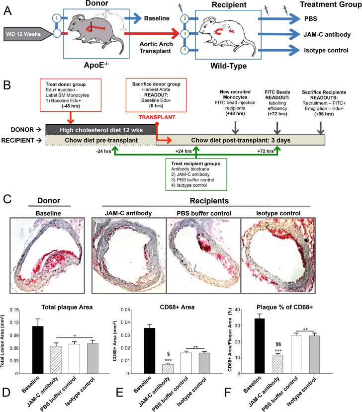
Fig 1. Ab blockade of JAM-C reduces plaques in a transplantation model of atherosclerosis.
(A) Scheme illustrating how atherosclerotic arches are generated in ApoE-/- donor mice preceding transplantation into treated or untreated normolipidemic WT mice. (B) Timeline of treatment and monocyte tracing in the aortic arches to establish recruitment and emigration profiles. Aortic arches were harvested from donor ApoE-/- mice (baseline) and transplanted or not into WT recipient mice treated with PBS buffer control, anti-JAM-C (JAM-C antibody) or an isotype control antibody (isotype control). Tissue sections were stained for CD68+ and visualized with Vector Red substrate. (C) Representative images from each group are shown. Morphometrics were analyzed using the ImageProPlus7 program with at least 2 measured areas per slide. To account for changes in the axial length of the arch, 4–7 indexes of sections were taken per arch, and 1 slide from each index was analyzed so that at least 4 slides were analyzed for each aortic arch vessel, and the mean value was used as the summary parameter. (D) Total plaque area, (E) CD68+ area, and (F) CD68+ as a percentage of the total plaque area were used as parameters of the lesion morphometrics. Data are presented as the mean ±SEM (N = 6). P values marked *—*** were calculated compared to baseline measurements. Both § and §§ = p<0.01 were compared to WT and IgG recipients.

