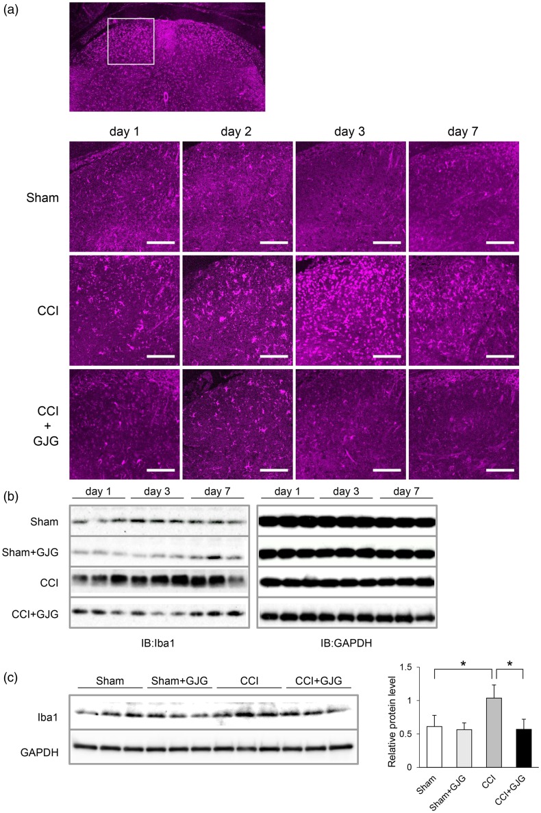
Figure 3.
GJG inhibited the expression of Iba1, a microglial activation marker, in the ipsilateral dorsal horn. (a) Immunohistochemical analysis of Iba1 from day 1 to day 7 post-operation. Confocal images were taken at 40 × magnification. Magenta fluorescence indicates Iba1-positive cells. Scale bar = 100 µm. (b) Results of Western blotting analysis of Iba1 from day 1 to day 7 post-operation. GAPDH is shown as a loading control. (c) Quantification of Western blotting analysis of Iba1 in Sham, GJG-treated Sham, CCI, and GJG-treated CCI groups on day 3 post-operation. The left blot was densitometrically analyzed, and the ratio of Iba1 immunoreactivity to GAPDH immunoreactivity was determined. Each data point represents the mean ± SD. n = 3. Data were analyzed using Dunnett’s test (*p < 0.05 indicates a significant difference between the indicated groups).

