Figure 10.
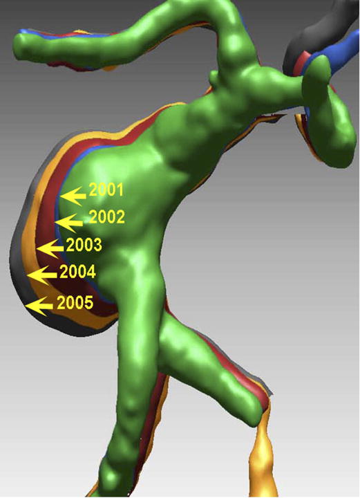
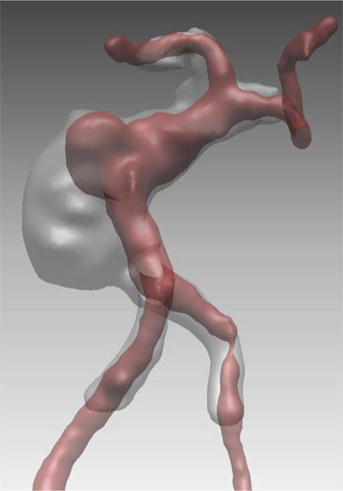
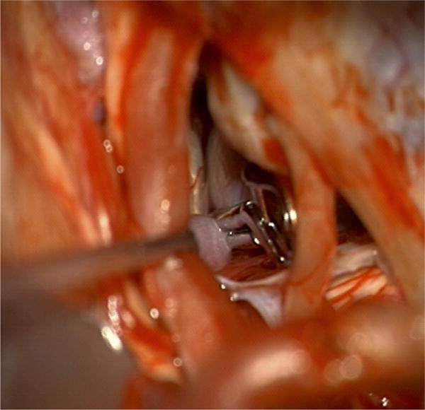
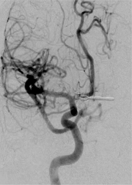
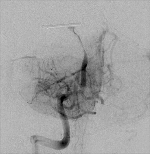
Case example of Phase 3 surgical management. (A) The 44-year-old man was followed with CE-MRA for 4 years with progressive aneurysm enlargement. (B) He then suffered a pontine stroke from acute thrombosis of the inferior portion of the aneurysm lumen, as shown on superimposed pre- (gray) and post-stroke luminal aneurysm volumes. (C) He underwent MCA-PCA bypass and distal clip occlusion of the aneurysm. (D) Postoperative angiography showed good perfusion of the basilar quadrifurcation through the bypass (right ICA angiogram, anteroposterior view) and (E) thrombosis of the remaining aneurysm lumen with anterograde flow up to the clip (right vertebral artery angiogram, anteroposterior view). The patient experienced a minor pontine perforator infarct resulting in a decrease in mRS score from 2 to 3, but he has required no further treatment in the subsequent 7 years and lives with some minor assistance.
