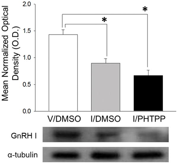
Figure 2. PHTPP Does Not Reverse Hypoglycemic Suppression of Rostral Preoptic Area Gonadotropin-Releasing Hormone (GnRH) GnRH-I Precursor Protein Expression.

GnRH-I precursor Western blot analysis was performed on rostral preoptic area (rPO) tissue obtained by calibrated hollow needle micropunch dissection 2 hr after sc injection of V (group 1) or I (groups 2 and 3). Twenty min before injections, animals were pretreated by delivery of DMSO alone (groups 1 and 2) or DMSO containing PHTPP (group 3) to the CV4. For each treatment group, lysate aliquots from individual subjects were combined to create four individual pools for Western blot analysis. Bars represent mean normalized GnRH-I precursor protein O.D. measures ± S.E.M. for V/DMSO (white bar), I/DMSO (gray bar), and I/PHTPP (black bar) groups. Typical GnRH-I and α-tubulin Western immunoblots are shown below the graph. *p <0.05, versus V/DMSO.
