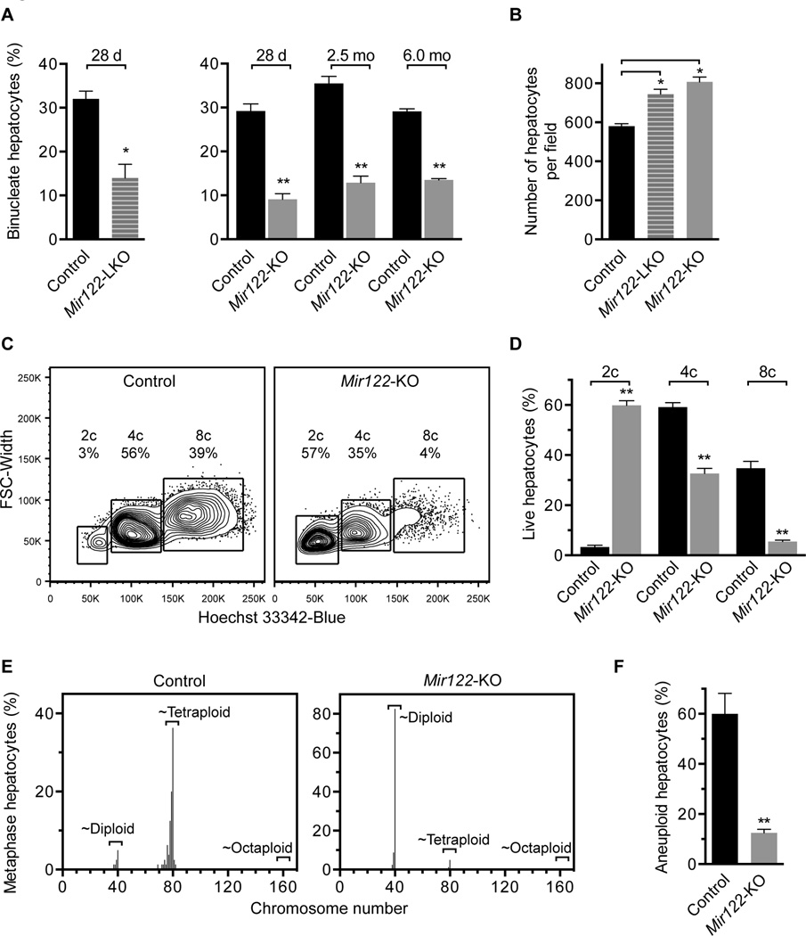
Fig. 3. Mir122-deficient livers are enriched for diploid, euploid hepatocytes.
(A,B) Livers from control, Mir122-LKO and Mir122-KO livers were stained with phalloidin and Hoechst 33342, as shown in Supporting Fig. 2A (n = 4–5 mice/age/genotype). The percentage of binucleate hepatocytes was reduced 2–3-fold at 28d (LKO and KO), 2.5mo (KO) and 6.0mo (KO) (A). 28d-old Mir122-LKO and Mir122-KO livers had 20–25% more hepatocytes per 150× field-of-view (B). (C,D) Single cell suspensions of hepatocytes were stained with a viability marker and Hoechst 33342, allowing identification of live cells with 2c, 4c and 8c nuclear content, corresponding to diploid, tetraploid and octaploid hepatocytes, respectively. Representative ploidy profiles are shown (C) and the percentage of each population quantified (n = 4; 2.5mo) (D). (E,F) Metaphase cytogenetic analysis revealed skewed chromosome counts (hypo-diploid, hypo-tetraploid and hyper-tetraploid) in control hepatocytes but not Mir122-KO hepatocytes (n = 4) (E). The overall degree of aneuploidy in Mir122-KO hepatocytes was reduced 4–5-fold compared to controls (F). See Supporting Fig. 4 individual karyotypes. * P ≤ 0.03, ** P ≤ 0.0001. Graphs show mean ± SEM.

