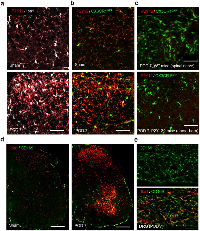
Figure 2. No monocyte infiltration in the spinal dorsal horn by P2Y12 and CD169 immunostaining.
(a) Representative confocal images of double immunostaining showing co-localization of P2Y12 (red) and Iba1 (white) signals in the spinal cord dorsal horn in sham and POD 7 following SNT in WT mice (n=4 per group). Scale bar is 50 μm. (b) Representative confocal images of P2Y12 staining in the spinal dorsal horn showing CX3CR1GFP positive (green) microglia are also P2Y12+ (red) in sham and POD 7 after SNT in CX3CR1GFP/+ mice (n=4 per group). Scale bar is 50 μm. (c) No P2Y12+ cells were found in the damaged nerve stump in CX3CR1GFP/+ mice and no P2Y12 immunostaining was found in the spinal dorsal horn of P2Y12−/− mice at POD 7 after SNT (n=4 per group). Scale bar is 50 μm. (d) Representative confocal images of double immunostaining showing co-localization of CD169 (green) and Iba1 (red) signals in the ipsilateral spinal cord at POD 7 in WT mice (n=2–3 mice per group). Scale bar is 100 μm. Note the obvious appearance of CD169+ cells (green) in the ipsilateral VH, but not DH. (e) CD169+ cell infiltration was also found in L4 dorsal root ganglia (DRG) at POD 7 in WT mice (n = 2–3 mice per group). Scale bar is 50 μm.

