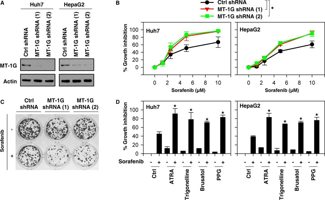
Figure 3. Suppression of metallothionein (MT)-1G expression enhances sorafenib sensitivity.
(A) Western blot analysis of MT-1G expression in indicated MT-1G knockdown hepatocellular carcinoma (HCC) cells. (B) Indicated MT-1G knockdown HCC cells were treated with sorafenib (1.25–10 µM) for 24 hours and cell viabilities were assayed (n=3, *p < 0.05). (C) Clonogenic cell survival assay. Indicated Huh7 cells were treated with sorafenib (5 µM) for 24 h, then 1,000 cells were plated into 24-well plates. Colonies were visualized by crystal violet staining two weeks later. (D) Indicated HCC cells were treated with sorafenib (5 µM) with or without all-trans retinoic acid (ATRA) (1 µM), trigonelline (0.5 µM), brusatol (40 nM), or propargylglycine (PPG) (2 mM) for 24 hours and cell viability was assayed (n=3, *p < 0.05 versus control group).

