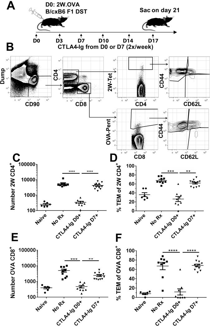
Figure 3. Endogenous antigen-specific CD4+ and CD8+T cell expansion is not reversed by delayed CTLA4-Ig treatment.
A, Diagram of experimental protocol. C57BL/6 mice were sensitized with 20×106 splenocytes from 2W-OVA B/6×B/c F1 mice. Recipients were treated biweekly with 250μg CTLA4-Ig 2x/week starting at either day 0 or 7; sensitized and untreated (No Rx), or naïve mice served as controls. B, Example gating strategy of sensitized C57BL/6 mouse. Combined spleens and axial, brachial, and inguinal LNs were stained and gated on Dump−CD90+ T cells, and separated into CD4+ and CD8+ gates. CD4+ and CD8+ T cells were examined for I-Ab:2W-binding (2W) or H-2Kb:OVA-binding (OVA), and for CD44 and CD62L expression. C&D, The total number of 2W CD4+ T cells, and CD44hiCD62Llo effectors among 2W cells is shown. E&F, The total number of OVA CD8+ T cells, and CD44hiCD62Llo effectors among OVA cells is shown. Data from 4 independent experiments (N=6-10/group) are presented; and statistically significant differences are indicated (**p<0.01, ***p<0.005, ****p<0.001).

