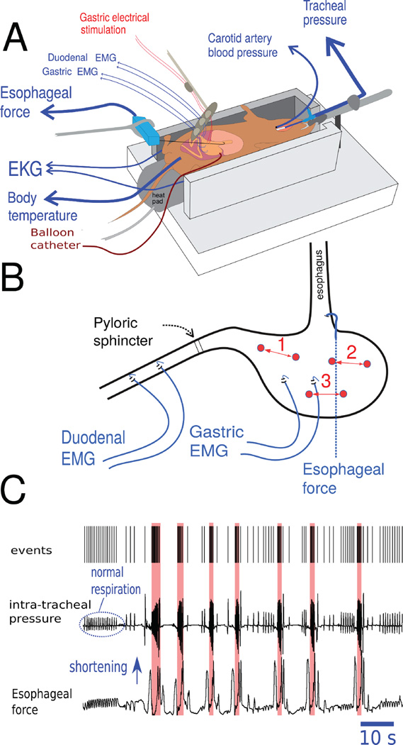
Figure 1.
Musk shrew in vivo anesthetized preparation. (A) Position of the recording electrodes, gastric electrical stimulation (GES) electrodes, and placement of the gastric balloon catheter for gastric distension. (B) GES electrodes were placed at three sites for the different studies, 1 = antrum, 2 = fundus, and 3 = body. Site 1 was used in Studies 1 to 3 and all three sites were used in Study 4. See Horn et al. (Fig. 7 in reference 16) for an anatomical image of the musk shrew stomach. (C) A representative example of detection of seven emetic episodes using the Kleinberg algorithm for burst detection (35). Detection of emetic episodes (red shading) was based on the sharp rise in frequency of respiration “events” in the top tracing. Apnea will often precede emesis and changes in esophageal force are shown as an additional indication of emesis (e.g., 32, 37).

