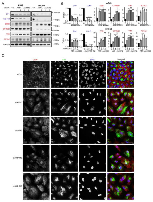
Figure 4. AMH and AMHR2 promote epithelial identity in NSCLC cells.
(A, B) Representative Western blots (A) and quantification (B) normalized to GAPDH loading controls indicating expression of proteins associated with an epithelial (ZO1 and CDH1) or a mesenchymal (ZEB1, CTNNB1, VIM and ACTA2) state, 48 hours after siRNA depletion of AMH and AMHR2. (C) Immunofluorescence visualization of expression of E-cadherin (CDH1), vimentin (VIM), or DNA (DAPI) 48 hours after siRNA depletion of AMH or AMHR2, or GL2 control (siCtrl). Scale bar, 30 μm. *, p<0.05; **, p<0.01; ***, p<0.001. Data are presented as mean ± SEM. See also Figure S5.

