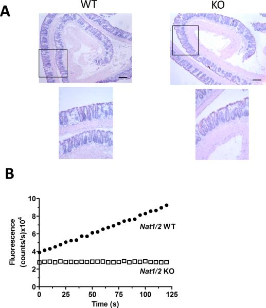
FIGURE 1.
Appearance of colonic mucosa and 5-ASA metabolism in Nat1/2 KO mice. (A) Representative images of hematoxylin and eosin stained distal colon in Nat1/2 WT and KO mice. Low resolution (4x) and high resolution (10x) of area indicated by rectangle in low resolution image. Bar = 0.2 mm (B) Representative traces of NAT enzyme activity show fluorescence (Ac-5-ASA: 312 nm excitation/ 437 nm emission) over 120 seconds of Nat1/2 WT (●) or Nat1/2 KO (□) mice distal colon homogenate immediately after the addition of 5-ASA and AcCoA.

