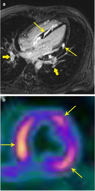
Fig. 12.

Sarcoidosis. a 32-year-old male admitted after a syncopal episode while shoveling snow. He was diagnosed with pulmonary sarcoidosis by transbronchial biopsy. Four-chamber delayed enhancement MRI shows patchy, diffuse areas of subepicardial as well as transmural areas of delayed post-contrast enhancement involving both ventricles (thin arrows). Bilateral hilar adenopathy was also seen (thick arrows). b FDG- PET after a fatty diet shows patchy areas of high uptake in the myocardium indicating areas of active inflammation
