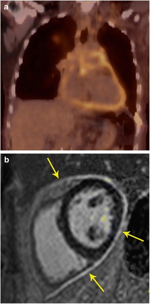
Fig. 14.

Pericarditis. a Coronal FDG PET/CT in a patient showed intense uptake in the pericardium. b Short-axis delayed enhancement MRI shows diffuse circumferential delayed pericardial enhancement (arrows), which is consistent with pericardial inflammation. There was also pericardial thickening in black blood images (not shown here)
