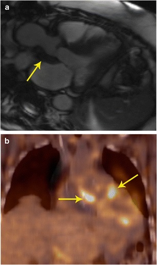
Fig. 15.

Perivalvular abscess. a Three-chamber Cine SSFP MRI image in a patient with history of bioprosthetic aortic valve replacement who presented with acute onset chest pain shows a soft-tissue intensity lesion surrounding the aortic valve and the aortic root (arrow). Contrast was not administered due to severe renal dysfunction. b Coronal FDG PET/CT in the same patient shows intense uptake along the lesion, which indicates that this is active inflammation/infection. Follow up MRI obtained two days later showed liquefaction and formation of abscess. This was treated surgically
