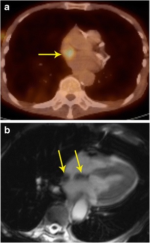
Fig. 5.

Localization of abnormal activity. a Axial FDG-PET/CT image shows a focus of high uptake adjacent to the SVC, which extended inferiorly. b Four-chamber SSFP MR image through the heart shows lipomatous hypertrophy of the interatrial septum (arrows), with sparing of fossa ovalis. This corresponds to the area of hypermetabolism in PET/CT. Although a benign lesion, this can occasionally show uptake in FDG-PET
