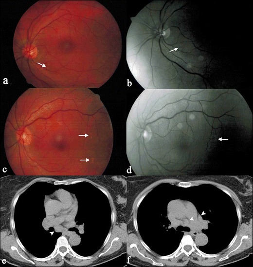
Fig. 20.

A 62-year-old woman with retinal involvement. Patient complained a left superior palpebral neoformation, which was subsequently biopsied: histological examination revealed the presence of a sarcoid nodule. She also referred to light flashes and blurred vision. Digital retinal angiography scan shows “punched-out” choroidoretinal lesions (white arrows on Fig. 20a–d). Chest CT (Fig. 20e and f) demonstrates mediastinal nodal enlargement, suggesting sarcoidosis in stage I
