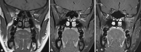
Fig. 10.

a A 35-year-old female presenting with painful opthalmoplegia of the left eye, diagnosed as IPT. Coronal T1-weighted MR image in the region of the orbital apex shows hypointense infiltrative soft tissue in the left orbital apex (straight arrow) extending inferiorly into the left PPF (curved arrow) via the left IOF (asterisk). Note the normal hyperintense fat within the right PPF (arrowhead). b Corresponding coronal contrast-enhanced FS T1W MR image in the same patient shows enhancing soft tissue in the left orbital apex (straight arrow) extending inferiorly into the left PPF (curved arrow) via the left IOF (asterisk). c Coronal contrast-enhanced FS TIW MR image in the same patient shows enhancement in the enlarged left foramen rotundum (arrow) in keeping with retrograde disease spread via the left V2 nerve. Note the normal right foramen rotundum (arrowhead)
