Fig. 3.

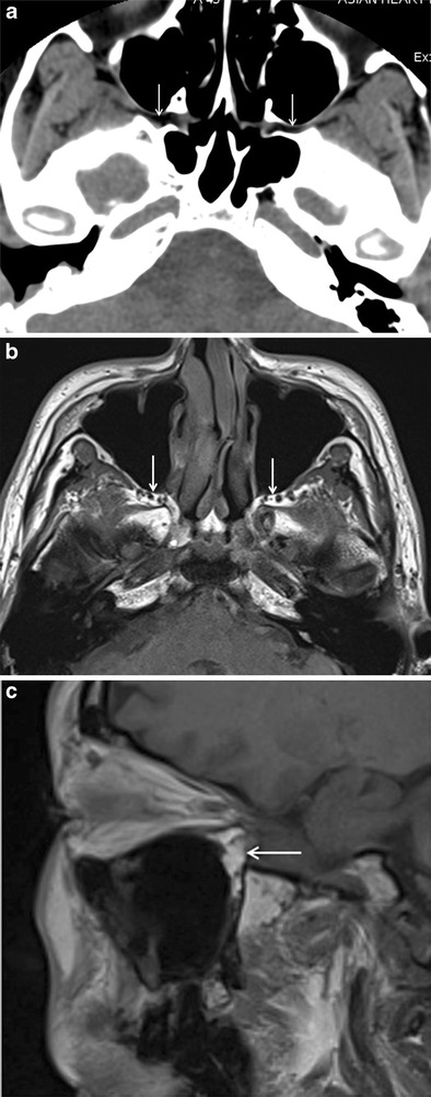
a Axial NECT image (soft-tissue window) showing normal hypodense appearance of bilateral fat-filled PPF (arrows). b and c Axial T1W MR image and sagittal T1W MR image showing hyperintense appearance of normal fat-filled PPF (arrows)

a Axial NECT image (soft-tissue window) showing normal hypodense appearance of bilateral fat-filled PPF (arrows). b and c Axial T1W MR image and sagittal T1W MR image showing hyperintense appearance of normal fat-filled PPF (arrows)