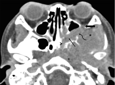
Fig. 6.

RMS of the left ITF in a 5-year-old boy. Axial CECT image shows an enhancing mass in the left ITF (asterisk) extending via the PMF (curved arrow) into the left PPF (thin straight arrow). The tumour extends via the IOF (thick arrow) into the left orbit. The left Vidian canal (arrowhead) is also involved
