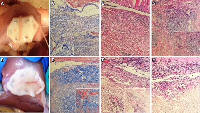
Fig. 3.
Mesh neovascularization, all images taken at 10× (large) or 20× (inset) magnification. Significant differences in neovascularization of implanted mesh were noted between experimental groups at the gross level (A - control, B - PRP+). Histologic analysis of Masson’s trichrome stained specimens confirms this effect, with significant difference in both the size and number of neovessels (orange-red) and depth of penetration into the mesh (blue) of control (C) versus PRP-treated (D) samples. Additional differences were noted in degree and depth of tissue ingrowth and immune cell reaction as seen in H&E stained specimens. Control samples displayed less ingrowth (E) and more chronic inflammatory infiltrate (G) compared to PRP-treated samples (F, H)

