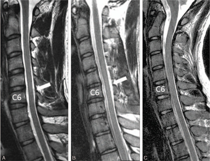
Figure 1.

(A) Sagittal T2-weighted cervical magnetic resonance imaging (MRI) of the spinal cord atrophied at the C5 to C6 vertebral body levels (white arrow) in a 22-year-old man with progressive distal atrophy of the right hand for 2 years. (B) Sagittal T2-weighted cervical MRI of the spinal cord atrophied at the C6 vertebral level (white arrow) in a 17-year-old man with progressive distal atrophy of the right hand for 6 months. (C) Sagittal T2-weighted cervical MRI of the spinal cord without cord atrophy in a 19-year-old man with progressive distal atrophy of the right hand for 2 years.
