
Figure 6.
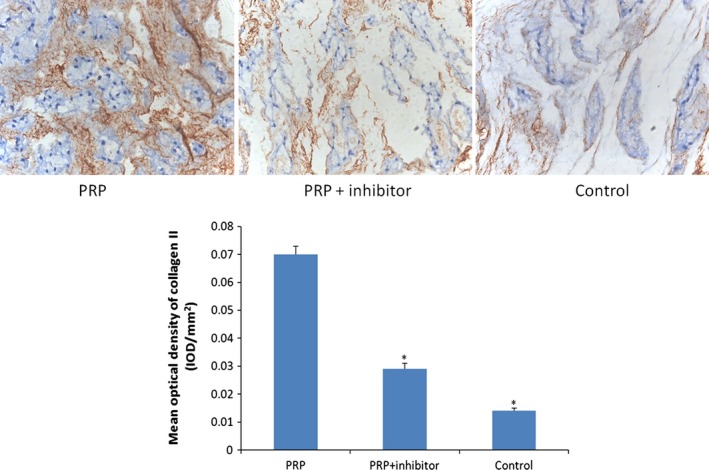
Collagen II immunohistochemical staining (×200). Collagen II, which provides the extracellular matrix framework of cartilage, was detected in PRP group at 8 weeks. Quantification of immunohistochemical staining showed that the PRP‐treated discs had significantly higher mean optical density than the transforming growth factor‐β1 inhibitor–treated and control groups (*P < 0.05 compared to the PRP group).