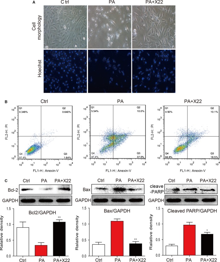
Figure 4.

X22 attenuated PA‐induced cell apoptosis in H9c2 cells. H9c2 cells were pretreated with X22 (20 μM) for 1 hr and then incubated with PA (500 μM) for 18 hrs. (A) Representative images for cell morphology analysis were obtained using light microscopy and Hoechst immunofluorescence staining. (B) Representative plots of flow cytometry with annexinV/PI. (C) The Western blot analysis for apoptotic proteins expression of Bax, Bcl‐2 and cleaved‐PARP in H9c2 cells (n = 3 for each experiment; *P < 0.05, **P < 0.01).
