Figure 6.

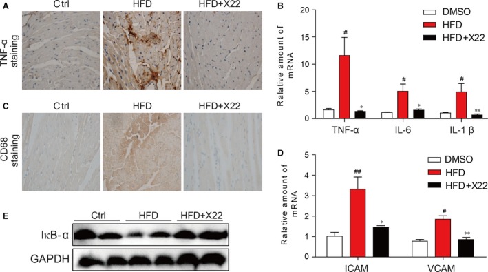
X22 attenuated HFD‐induced inflammation in HFD‐fed rats. (A) Representative images for immunohistochemical staining for TNF‐α accumulation in the formalin‐fixed myocardial tissues (400× magnification). (B) The mRNA expression of the inflammation markers IL‐6, IL‐1β and TNF‐α in the myocardial tissues. (C) Representative images for immunohistochemical staining for CD68 accumulation in the formalin‐fixed myocardial tissues (400× magnification). (D) The mRNA expression of the adhesion markers VCAM‐1 and ICAM‐1 in the myocardial tissues. ICAM‐1: intercellular adhesion molecule‐1; VCAM‐1: vascular cell adhesion molecule‐1. (E) The Western blot analysis for the protein expression of IκB‐α in the myocardial tissues (#, versus DMSO samples; *, versus HFD samples; # and *P < 0.05, ## and **P < 0.01; n = 7 per group).
