Figure 7.

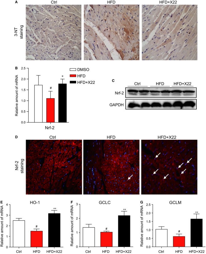
X22 attenuated HFD‐induced myocardial oxidative stress in HFD‐fed rats. (A) Representative images for immunohistochemical staining of 3‐NT accumulation in the formalin‐fixed myocardial tissues (400× magnification). (B) The mRNA expression of the anti‐oxidative markers Nrf‐2 in the myocardial tissues was determined by real‐time qPCR assay. (C) The Western blot analysis for the protein expression of Nrf‐2 in the myocardial tissues. (D) Representative images for immunohistochemical staining of Nrf2 and DAPI in the formalin‐fixed myocardial tissues (200× magnification). (E–G) The mRNA expression of the anti‐oxidative genes HO‐1, GCLC and GCLM in the heart tissues was determined by real‐time qPCR assay (*, versus HFD group; #, versus Ctrl group; * and # P < 0.05; ** and ## P < 0.01; n = 7 per group).
