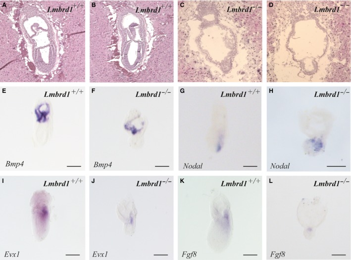
Figure 3.

Pre‐gastrulation markers are absent in Lmbrd1 −/−‐embryos. (A–D) Sagital sections of Lmbrd1 +/+‐embryos and Lmbrd1 −/−‐embryos followed by haematoxylin and eosin staining. Lmbrd1 −/−‐embryos exhibit extraembryonal structures, whereas in contrast to Lmbrd1 +/+‐embryos the epiblast is composed of only two cell layers at E7.5. (E–L) Lateral view of Lmbrd1 +/+‐ and Lmbrd1 −/−‐embryos stained by whole mount in situ hybridization. (E and D) Lmbrd1 +/+‐ and Lmbrd1 −/−‐embryos show similar expression of Bmp4 in extraembryonic tissues. (G and H) Detection of Nodal expression in both Lmbrd1 +/+‐ and Lmbrd1 −/−‐embryos. (I and J) Evx1 is detectable in the dorsal part of Lmbrd1 +/+‐embryos (I), whereas low levels of Evx1 are present in a restricted area in Lmbrd1 −/−‐embryos (J). (K and L) Fgf8 is expressed in the dorsal part of Lmbrd1 +/+‐embryos (K), whereas Fgf8 is partially expressed in Lmbrd1 −/−‐embryos (L). Scale bars represent 100 μm. Representative embryos are shown (n = 5).
