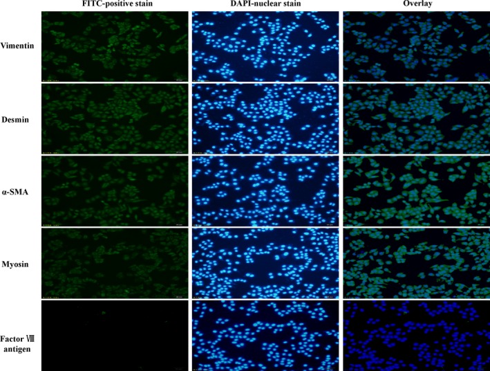
Figure 5.

Immunofluorescence photomicrographs of cultured cells. Immunofluorescence was performed on cultured cells using an inverted microscope (scale bar: 50 μm). The results showed that the cultured cells had positive staining for vimentin, desmin, α‐smooth muscle actin (α‐SMA) and myosin but negative staining for factor VIII antigen.
