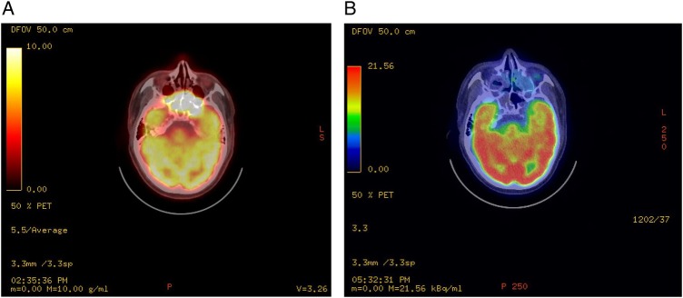
Figure 1.
(A) PET scan before treatment revealed very abnormal metabolically active soft tissue bone extending from the nasopharynx to maxillary antra, ethmoid, sphenoid, orbits and mid-cranial fossa. There is clear bone destruction of the sinuses walls and base of the skull. (B) Post-treatment PET scan performed 8 months later revealed complete disease remission of primary disease at the base of skull. There is no local metabolically active disease. PET, positron emission tomography.

