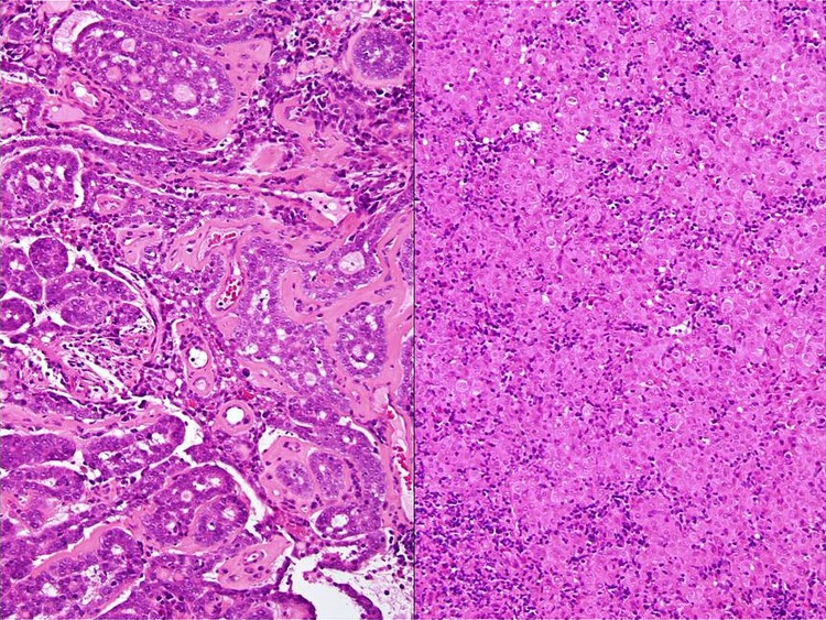
Figure 3.
Photomicrographs of histology tissue (image on the left) showing low-grade adenoid cystic carcinoma, tubulo-cribriform pattern with abundant eosinophilic basement membrane material, numerous pseudolumina and occasional true glandular lumina, contrasted against (image on the right) more poorly differentiated elements showing well-defined, pleomorphic, large epithelioid cells disposed in solid/insular architecture. The latter is accompanied by more conspicuous interstitial lymphoplasmacytic inflammatory cell infiltrate albeit with minimal intratumoural lymphocytosis proper (H&E; original magnifications ×10).

