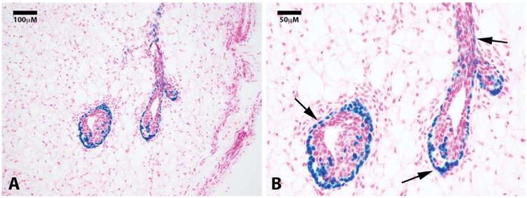
Figure 1.

Clonogenic demonstration of labeled mammary cap cells and progeny. Photomicrographs of a nulliparous gland 4 weeks after H253 implantation show positive lacZ staining in the cap cells of terminal end buds at 20× (A) and 40× (B) magnification. Sections were counterstained with nuclear fast red. Black arrows point to positive cells in the basal epithelial layers.
