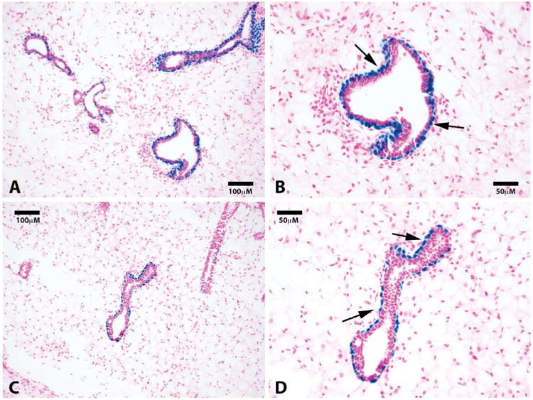
Figure 2.

Luminal cells along subtending ducts are derived from a separate lacZ negative progenitor. Representative photomicrographs of mammary ducts at 20× (A) and 40× (B) show positive staining for lacZ cells in the myoepithelial cells along the basal surface. No luminal lacZ positive cells are found. Sections were counterstained with nuclear fast red. Black arrows point to lacZ positive basal cells.
