FIGURE 4.
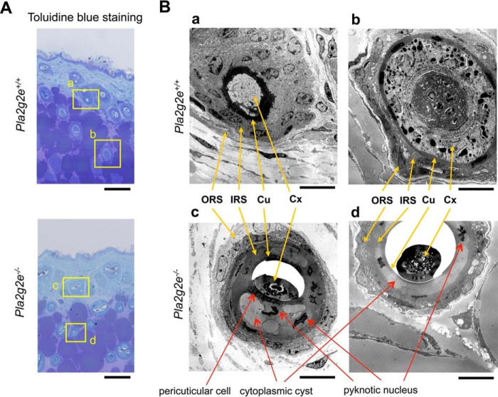
Transmission electron microscopy of Pla2g2e+/+ and Pla2g2e−/− skins. A, toluidine blue staining of Pla2g2e+/+ and Pla2g2e−/− skins. Boxed areas (panels a–d) are magnified in B. Bars, 100 μm. B, transmission electron microscopy of hair follicles in Pla2g2e+/+ (panels a and b) and Pla2g2e−/− (panels c and d) skins. Locations of ORS, IRS, cuticle (Cu), and cortex (Cx) are indicated by yellow arrows. Red arrows indicate abnormal features observed in Pla2g2e−/− mice (formation of cytoplasmic cysts and pyknotic nuclei in the IRS and the presence of pericuticular cells). In addition, the cuticle and cortex were unusually dissociated in Pla2g2e−/− mice. Bars, 10 μm.

