FIGURE 6.
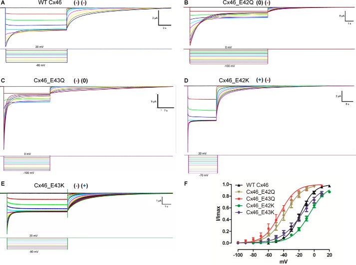
Determination of voltage dependence of the slow gate in WT and mutant Cx46 hemichannels. Oocytes maintained at a holding potential of +20 mV (WT Cx46, Cx46_E42K, and Cx46_E43K) or 0 mV (Cx46_E42Q and Cx46_E43Q) were subjected to hyperpolarizing pulses from 20 to −60 mV (WT Cx46), from 0 to −110 mV (Cx46_E42Q), from 0 to −100 mV (Cx46_E43Q), from 20 to −70 mV (Cx46_E42K), or from 20 to −90 mV (Cx46_E43K) and returned to holding potential. Hemichannel currents recorded in oocytes expressing Cx46 (A), Cx46_E42Q (B), Cx46_E43Q (C), Cx46_E42K (D), and (Cx46_E43K) (E) mutants. F, graph depicting the I(V)/Imax relation of WT Cx46 (black), Cx46_E42Q (brown), Cx_E43Q (red), Cx_E42K (green), and Cx_E43K (blue) mutants. The solid lines represent the fitting of the data to Equation 7 and using the mean values in Table 4. Current traces and corresponding voltage traces have the same color.

