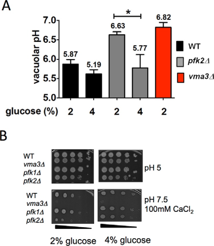
FIGURE 2.

Metabolic reactivation is defective in pfk2Δ cells. A, the acidic vacuolar pH is restored in 4% glucose. Wild type, pfk2Δ, and vma3Δ cells were cultured to mid-log phase in YEP containing 2% or 4% glucose. The cells were harvested and stained with 50 μm BCECF-AM for 30 min at 30 °C. The ratio of fluorescent emission (535 nm) excited at 490 and 450 nm was measured to quantitatively assess vacuolar pH. The average fluorescence over 6 min was compared with a standard curve to generate absolute pH values. Data are presented as average pH values from three independent experiments, and error bars are standard deviation. Statistically significant differences (*, p < 0.05) were determined by two-tailed unpaired t test. B, the vma− growth phenotype is rescued in 4% glucose. Cells were cultured to mid-log phase in 2% or 4% glucose, and 10-fold serial dilutions were stamped onto YEP plates containing 2% or 4% glucose adjusted to pH 5.0 and pH 7.5 plus 100 mm CaCl2. Cell growth was monitored for 72 h at 37 °C. Shown are representative plates of triplicates.
