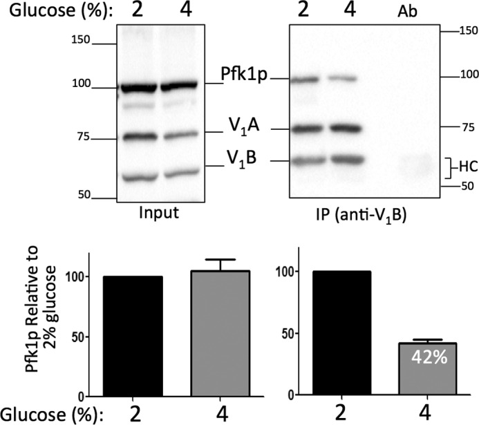
FIGURE 6.

Pfk1p subunit binding to V-ATPase decreases in 4% glucose. Overnight mid-log phase cultures (optical density of 0.8–1.0 A600/ml) were lysed, and V-ATPase complexes were immunoprecipitated with anti-A monoclonal antibody. Immunoprecipitated protein (IP) and total lysate protein (Input) were loaded on 10% SDS-PAGE gels. Pfk1p and V-ATPase (V1 subunits A and B) were detected by immunoblots using, respectively, anti-PFK-1 polyclonal antibodies and anti-B and anti-A monoclonal antibodies and horseradish peroxidase secondary antibodies. Ab, antibody alone; HC, antibody heavy chain. Protein markers are 150, 100, 75, and 50 kDa. A representative gel is shown (top panel). Gels from two independent experiments were scanned using a Bio-Rad ChemiDoc XRS+, and data were analyzed using Multi Gauge V3.0 and GraphPad Prism 5 software. Data were expressed as –fold increase Pfk1p:V1 subunit ratio ± S.D. relative to the wild type (bottom panel).
