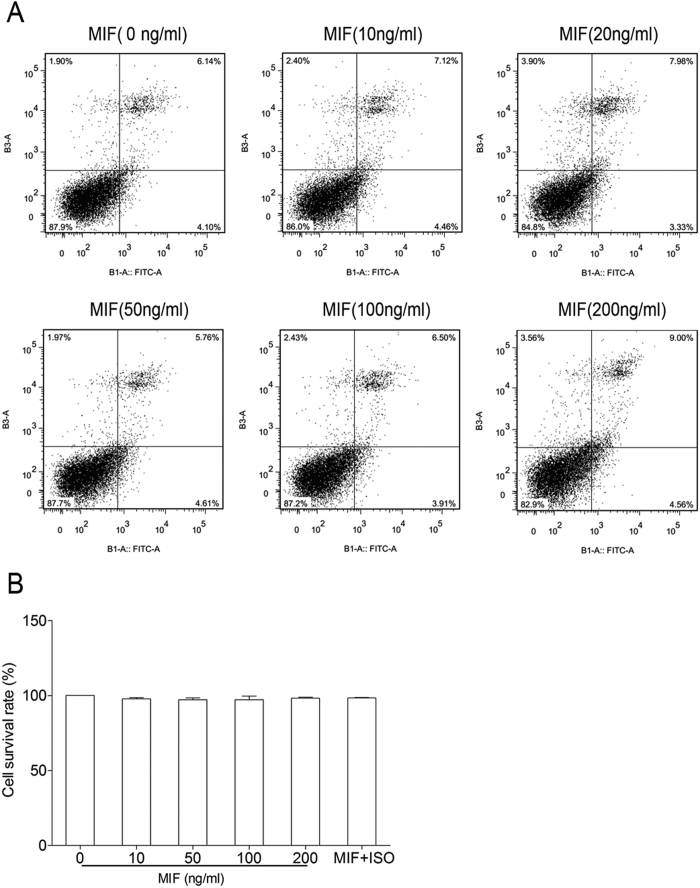
Figure 11. Effect of MIF on cell viability and apoptosis of RAW264.7 cells.
RAW264.7 cells were treated with different concentrations of MIF (0, 10, 20, 50, 100, 200 ng/ml) for 24 hrs. In experiment of ISO-1 treatment, RAW264.7 cells were pre-treated with 100 μg/ml ISO-1 for 30 min, following stimulated with 100 ng/ml MIF for 24 hrs. Flow cytometry assay was used to examine the apoptosis of cells and Cell Counting Kit-8 was used to measure cell viability. Bar graphs represent the mean ± SEM of three independent experiments.

