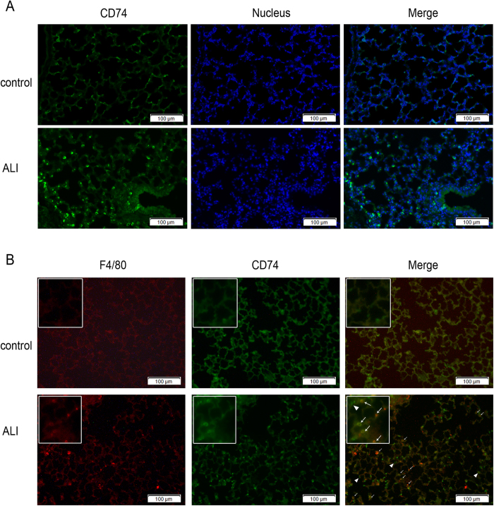
Figure 4. Immunofluorescence staining of CD74 in lungs.
Immunofluorescence examination was performed for CD74 in control mouse lung tissue and lipopolysaccharide induced acute lung injury model. Increased surface CD74 expression (green) in lung tissue of acute lung injury was observed compared with control (A,B). Surface CD74-positive cells were observed mainly on the alveolar septa, and colocalize with F4/80-positive cells (macrophage cells; red) (B). Arrows, positive staining of macrophage cells; arrowhead, positive type II alveolar epithelial cells. Scale bar represents 100 μm.

