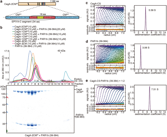
Figure 2. Two PAR1b can simultaneously bind to CagA.
(a) Schematic diagram of CagA-2CMW and a blow-up of the region containing the CM motifs. CMW1 resides in the 34-amino-acid EPIYA-C segment and CMW2 flanks the segment on the C-terminal side. The two CM sequences are 18 amino acid residues apart. (b) Results from the size-exclusion chromatography of CagA-PAR1b (39–364) complexes analysed on Superdex 200 10/300 GL using HBS-P as running buffer (top). Fractionated CagA-2CM + PAR1b (39–364) complex was further resolved on SDS-PAGE gel and stained by CBB (bottom). (c–e) Analytical ultracentrifugation of 5.5 μM CagA-C1164S (CagA-CS) (c), 11 μM PAR1b (39–364) (d), and 5.5 μM CagA-CS + 11 μM PAR1b (39–364) (e). Raw absorbance distributions, the best-fit model (left), and the continuous sedimentation distributions c(s) (right) calculated by the SEDFIT program.

