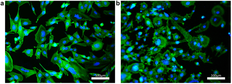
Figure 3.
Fluorescent images of breast cancer cell line MDA-MB 231 showing the morphological changes of cells grown on control glass (a) and horizontal GAIN scaffolds (b), with evident increased number of microspikes and initial stages of cytoplasm infiltration into the scaffolds supposing high membrane activity.

