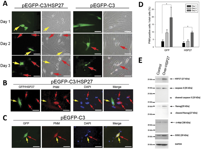
Figure 3. Physiological effects of over-expression of HSP27 in PDMCs.
The PDMCs were induced with IBMX for 6 h and then transfected with the pEGFP-C3 or pEGFP-C3/HSP27 plasmids, as indicated. (A) The cells were fixed and directly visualized using fluorescence microscopy at 1 day, 2 days and 3 days after transfection. The green cells show the expression of GFP. The red arrows indicate the neuron-like cells, and yellow arrows indicate the GFP-expressing cells. (B) Cells from each transfection were fixed and probed with Pan-Neuronal Marker (PNM) antibody for visualizing neuronal cells. The GFP/HSP27-transfected cells showed green signal alone, without co-localization with PNM-positive cells. (C) Without HSP27 overexpression, there was partial colocalization of the GFP and PNM signals. (Magnification = 200x). (D) The transfected cells in each condition were probed with PNM antibody, and PNM fluorescence signals were recorded and quantified. The cells showing PNM immunofluorescence were counted as positive cells. The proportion of differentiated neurons was quantified as the positive PNM immunofluorescence staining divided by the DAPI-positive cells. (E) Immunoblots of PDMCs with HSP27 overexpression. The upregulated HSP27 protein expression levels were confirmed in HSP27-overexpressing cells (right column) compared with controls. HSP27-overexpressing PDMCs (indicated by arrowheads) showed no cleaved forms of caspase-3 or Nanog. Additionally, the cleaved forms of SOX2 and c-myc, two other stem cell markers, were absent in both experimental conditions. GAPDH was used as a loading control for immunoblotting.

