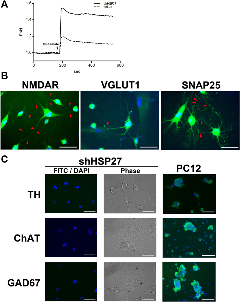
Figure 6. Knockdown of HSP27 directs PDMCs to differentiate into glutamatergic neurons.
(A) Neuronal function in induced, HSP27-silenced PDMCs was determined based on Ca2+ influx profiles. The arrow indicates the addition of glutamate (20 μM). The plot shows the higher-magnitude kinetic profile in HSP27-silenced PDMCs after glutamate addition compared with Luc-silenced cells. (B) Immunofluorescent analysis of shHSP27-infected PDMCs induced with 0.4 mM IBMX. The cells were stained with primary antibodies against the glutamatergic neuron markers NMDAR, VGLUT1 and SNAP25, followed by the appropriate FITC-conjugated secondary antibodies. The cell nuclei were counterstained with 4′,6-diamino-2-phenylindole (DAPI). The attachment of a dendrite from another cell (red arrow) and synaptic connections (red arrowhead) are indicated. (C) Induced neurons derived from HSP27-silenced cells were stained for a dopaminergic neuron marker, TH; a cholinergic neuron marker, ChAT; and a GABAergic neuron marker, GAD67. The FITC and DAPI images for each condition were merged. The phase contrast images of each condition are also shown to demonstrate the neuronal phenotype of each staining. PC12 cells were used as positive controls for each antibody. (Scale bar = 100 μm). Abbreviations: N-methyl-D-aspartate receptor, NMDAR; vesicular glutamate transporter 1, VGLUT1; synaptosomal-associated protein 25, SNAP25; tyrosine hydroxylase, TH; choline acetyltransferase, ChAT; and glutamic acid decarboxylase, GAD67.

