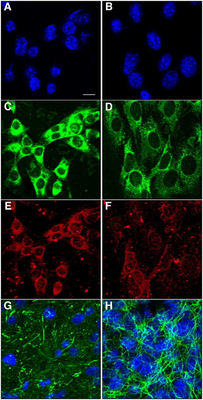
Figure 1. Immunocytochemical characterization of brain endothelial cells.
Wt (A,C,E) and Psen1−/− (B,D,F) brain endothelial cells were fixed with acetone/methanol and immunostained for laminin (C,D) and PECAM (CD31; E,F) along with a DAPI nuclear stain (A,B). Panels (G,H) show confocal images of Wt (G) and Psen1−/− (H) endothelial cells immunostained for fibronectin (green) with DAPI counterstaining (blue). Scale bar, 10 μm.

