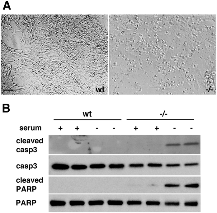
Figure 2. Serum starvation results in apoptosis of Psen1−/− endothelial cells.
(A) Brightfield micrographs of wt (left) and Psen1−/− cells (right) after serum starvation for 16 h. Note the rounded appearance of the Psen1−/− cells in contrast to the normal appearance of wt cells. Scale bar, 20 μm. (B) Western blot detection of cleaved and total caspase 3 (casp3) or PARP from cells grown for 16 h under serum containing (+) or serum starved (−) conditions. Note the presence of cleaved fragments of caspase 3 and PARP, both markers of apoptosis, in serum-starved Psen1−/− cells but not in Psen1−/− cells grown in serum or in wt cells maintained under either condition. The blots are representative of three independent experiments.

