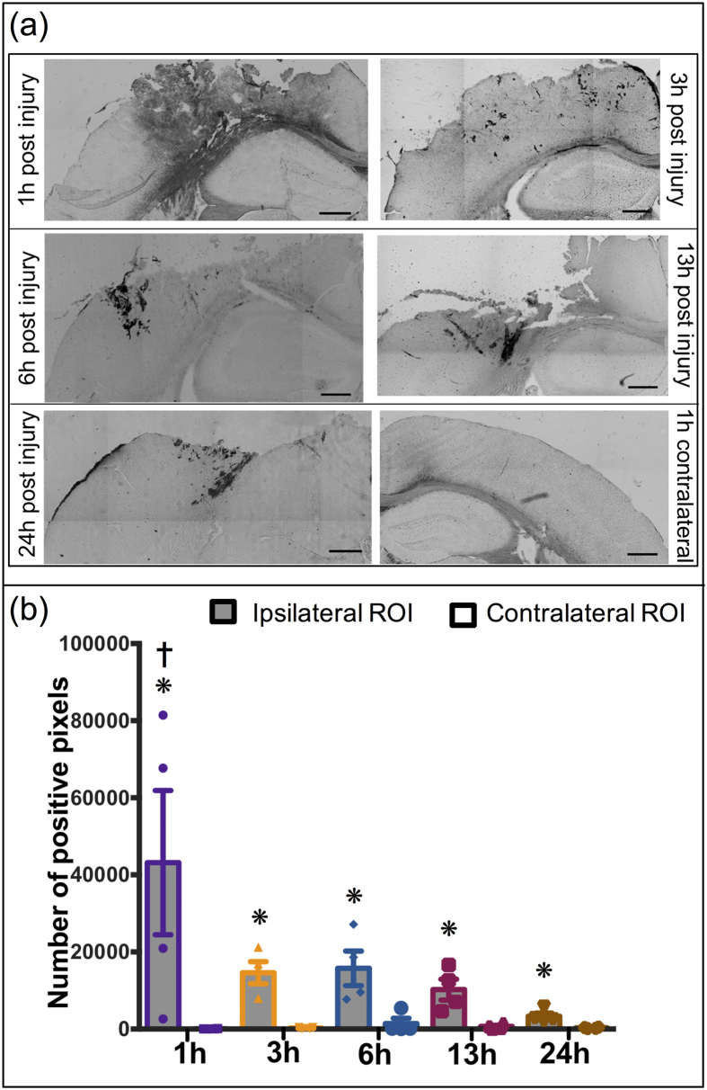
Figure 2.
HRP Extravasation after TBI: (a) Representative images of extravasation of HRP injured region after 1 h, 3 h, 6 h, 13 h and 24 h post injury (a–e); contralateral region 1 h post injury (f). (b) Quantitative analysis of HRP extravasation over time. *p < 0.05 compared to their respective contralateral ROI, Student’s t-test. †p < 0.05 compared to 13 h and 24 h ipsilateral ROI, Tukey’s post-hoc test. Error bars represents standard error of mean with n = 4 per group. Scale bar = 500 μm.

