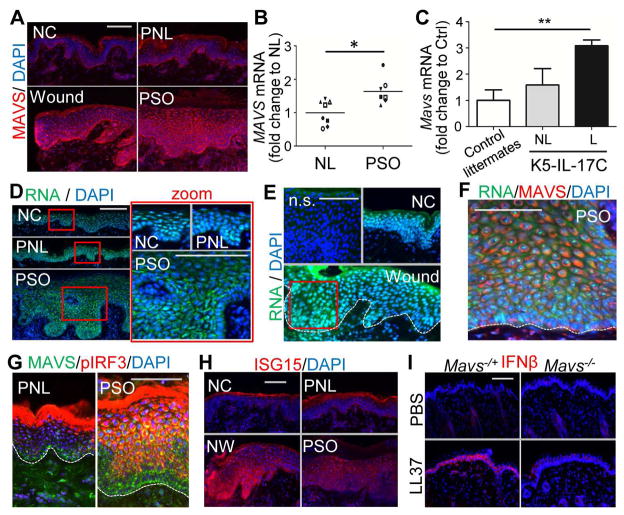
Fig. 7. Activation of MAVS-IRF3-IFNβ signaling in psoriatic and wounded skin epidermis.
(A) MAVS immunostaining of skin sections from normal control skin (NC), wounded (W), psoriasis non-lesional (PNL) or psoriasis lesional (PSO) skin as indicated. (B–C) Measurement of relative MAVS mRNA levels in (B) non-lesional (NL) and psoriasis lesional (PSO) skin (n=7~8/group), or (C) in non-lesional (NL) and lesional (L) of K5-IL-17C mouse skin compared with littermate control skin (n=4~5/group). (D–F) Human psoriatic or wounded skin sections were stained with RNA dye (green) and DAPI (D), or RNA, MAVS antibody and DAPI (E), or anti-MAVS and anti-pIRF3 antibodies (F), or ISG15 (G) as indicated. Zoom-in polyICtures highlighted in red box are shown on the right panel. White dashed line indicates the junction of the epidermis and dermis. (H) Representative image of IFNβ immunostaining in Mavs−/+ or Mavs−/− mouse skin injected intradermally with LL37. All scale bars = 100 μm.

