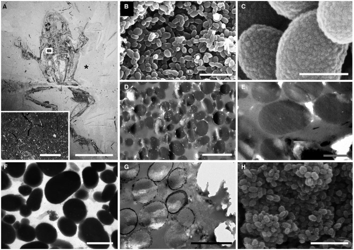
Figure 1.

Melanosome‐like microbodies in the Libros frogs. A, MNCN 63663; inset, detail of thin, dark brown carbonaceous film defining soft tissues in area indicated; asterisk indicates region of sediment analysed in Fig. 4E. B–H, scanning (B–C, H) and transmission (D–G) electron micrographs showing details of melanosome‐like microbodies in the brown layer. B–C, densely packed melanosome‐like microbodies (B) with detail of surface texture (C). D–F, unstained (D–E) and stained (F) TEM sections of microbodies showing uniform electron density; note internal vacuoles in microbodies in D. G–H, unstained sections of microbodies immediately adjacent to the phosphatized skin showing electron‐dense margin of calcium phosphate (G) and nanocrystalline surface texture (H). Scale bars represent: 50 mm (A); 1 mm (inset in A); 5 μm (B, F); 500 nm C, E–F); 1 μm (D); 2 μm (G).
