Fig. 2.

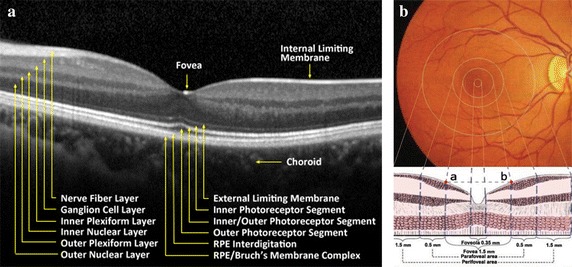
Diagram of normal retinal structure. a Normal retinal tissue layers image shot by OCT instrument. b Macular structure, including foveola, fovea, parafovea, perifovea regions

Diagram of normal retinal structure. a Normal retinal tissue layers image shot by OCT instrument. b Macular structure, including foveola, fovea, parafovea, perifovea regions