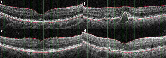
Fig. 9.

The results of retinal boundary extraction in OCT images. After the preprocessing of standardization and de-noising, the ILM and RPE boundaries are extracted based on intensity features, as shown in the red lines. The green lines represent the medical macular regional division boundaries. a–d are respectively corresponding to the four original images in Fig. 8
