
Fig. 5.
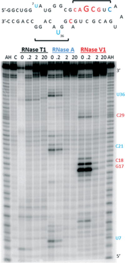
Top: Summary of RNase protection of RRE IIB with A6. Font size reflects levels of protection from RNase A (blue) and RNase VI (red), and brackets indicate regions of peptide: RNA contact; Bottom: Autoradiograph of footprinting experiment. AH is the alkaline hydrolysis ladder and C is the negative control (RRE IIB only, with no RNase or peptide).