Fig. 1.

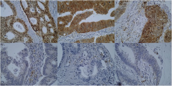
p62 immunohistochemistry. Examples of p62 immunostaining in colorectal cancers. The upper row shows three tumours with strong cytoplasmic immunostaining and the lower row demonstrates three tumours with negative p62 staining (×400 magnification). Note the positively stained of stromal cells/macrophages, which can be used as internal controls
